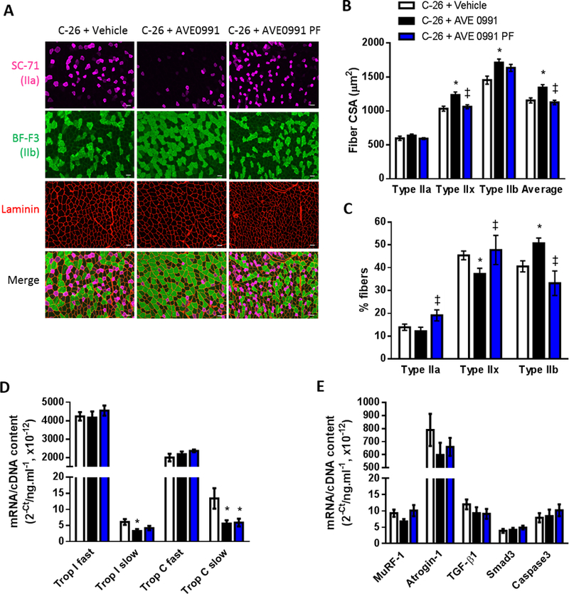
Figure 7.

High dose AVE0991 (15 mg/kg) induces TA muscle fiber hypertrophy in severely cachectic C-26 tumor-bearing mice fed ad libitum. (A) Representative images of TA muscle cross-sections reacted for laminin (red) to indicate all muscle fibers and myosin IIa (SC-71, pink) and myosin IIb (BF-F3, green) to indicate type IIa and type IIb fibers, respectively. Since mouse TA muscles do not express detectable levels of type I fibers, those fibers not reacting with SC-71 or BF-F3 (i.e. black fibers in the merge) were assumed to be type IIx fibers. Quantification of SC-71, BF-F3 and laminin based on reaction intensity facilitated determination of: (B) average fiber CSA and CSA of the type IIa, type IIx (non-reacting with SC-71 or BF-F3) and type IIb fibers; and (C) the proportion of type IIa, type IIx and type IIb fibers (one-way ANOVA; *P<0.05 vs. C-26+Vehicle; ‡P<0.05 vs. C-26+AVE0991; C-26+Vehicle, n=16; C-26+AVE0991, n=15; C-26+AVE0991 PF, n=7). Gene expression of (D) the fast and slow isoforms of troponin I and troponin C, (E) the ubiquitin ligases MuRF-1 and atrogin-1, TGF-β1, Smad3 and caspase3 in TA muscles from vehicle or AVE0991 treated C-26 tumor-bearing mice (one-way ANOVA; *P<0.05 vs. C-26+Vehicle; n=7–8. Using a 20× objective, total magnification for images in Fig. 7A is 126×. Scale bar = 50 µm.
