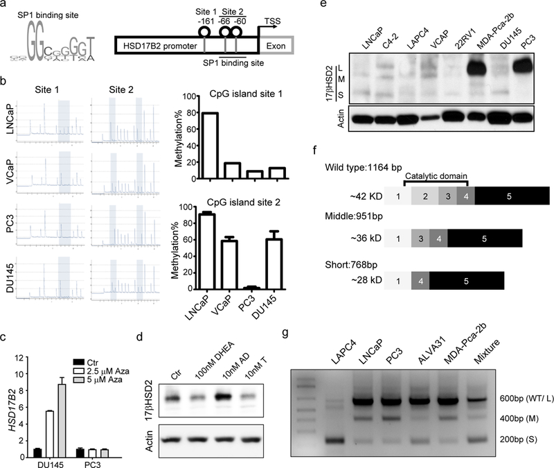
Figure 4. Multiple regulation mechanisms of HSD17B2 functional silencing.

(a) Schema of SP1 binding site and HSD17B2 promoter. (b) DNA methylation of HSD17B2 promoter in prostate cancer cell lines. (c) HSD17B2 expression after 5-azacytidine treatment. HSD17B2 expression was normalized to RPLPO. Basal expression of HSD17B2 in each cell lines was taken as 1. (d) 17βHSD2 protein abundance after androgen stimulation. PC3 cells were treated with indicated androgens for 8h before collection for western blot. (e) Endogenous 17βHSD2 expression in different cell lines. (f) Schema of different HSD17B2 isoforms. (g) Amplicon of different isoforms in prostate cancer cell lines. Primers located in HSD17B2 exon 1 and exon 4 were used for PCR.
