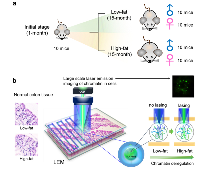
Fig. 1.
Conceptual illustrations of the experimental design and setup. a, Experimental animal model used in this work. We focused on the precursor of very early stage development of adenomas by using mouse colon tissues as a model system. Formalin fixed paraffin-embedded (FFPE) tissues from 50 mice were examined, including 10 1-month mice (5 males and 5 females), 40 15-month mice (20 males and 20 females with low-fat and high-fat dietary treatment). b, Conceptual illustration of a large-scale rapid scanning laser-emission microscopy (LEM) for studying cancer precursors, in which an FFPE tissue is sandwiched within a high-Q Fabry-Pérot (FP) cavity. Tissue thickness for all samples used in LEM was 10 μm. Excitation wavelength = 473 nm. Details can be found in Fig. 2. The left panel shows that no significant morphological difference is observed in the H&E images between a low-fat colon tissue and an “apparently normal” high-fat colon tissue. The right panel shows that significant differences can be seen in the LEM images (and other lasing characteristics) between the same two tissues in the left panel. For example, the high-fat tissue has a lower lasing threshold, and more lasing cells and stronger lasing emission under the same external excitation than the low-fat tissue. Those differences reflect the abnormal cell proliferation and chromatin deregulation (condensation) in the high-fat colon tissue, despite the absence of abnormal morphological changes in the H&E image.

