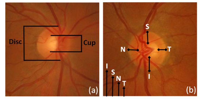
Fig. 1.
Example of findings used to detect glaucoma in color fundus images. (a) Quantification of the optic cup to disc ratio (CDR). The reduction of the optic nerve fibres (typically related with glaucoma) provokes optic disc cupping, central cup becomes larger, with respect to the optic disc (b) The neuroretinal rim usually follows a normal pattern (ISNT rule) where the inferior region is broader than the superior, broader than the nasal, and broader than the temporal region. The alteration of this pattern is a suspicious sign of glaucoma

