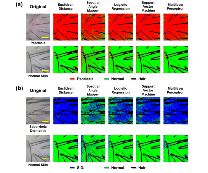
Fig. 6.
Spectral classification using the constructed models: (a) photographic and classified images of psoriasis (top) and normal skin regions (bottom) and (b) photographic and classified images of seborrheic dermatitis (SD)(top) and normal skin regions (bottom) on the scalp (field of view: 3.3 mm x 2.99 mm, pixel size: 760 x 651, and magnification: 4x).

