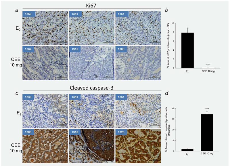
Figure 5.
Effects of E2 and CEE on cellular proliferation marker Ki67 and cleaved caspase-3 in mammary tumors from ACI rats. (a) Images of immunohistological staining of Ki67 of three sections from each group. (b) Percentage of area of Ki67 positive cells of all tumor sections examined. *****p = 0.000003. (c) Images of immunohistological staining of cleaved caspase-3 of three sections from each group. Animal ID number is shown in left upper corner of each panel. (b) Percentage of area of cleaved caspase-3 positive cells of all tumor sections examined. *****p = 0.0000003.

