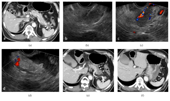
Figure 4.
A patient who developed bleeding with endoscopic ultrasound-guided fine needle biopsy (EUS-FNB) using a Franseen needle. (a) Contrast-enhanced Computed Tomography (CT) scan showing a 3-cm hypovascular mass in the pancreatic tail (arrow). (b) Insertion of the needle under EUS guidance. (c) Active bleeding from the needle tract right was noticed under color Doppler mode after the withdrawal of the needle. (d) Increased echo-free space between the pancreas and stomach was identified. ((e), (f)) Contrast-enhanced CT scan was performed immediately after EUS-FNB. Hyperdense fluid collection suggesting hematoma was observed between the pancreatic tail and the greater curvature of the stomach (arrow).

