Figure 1.

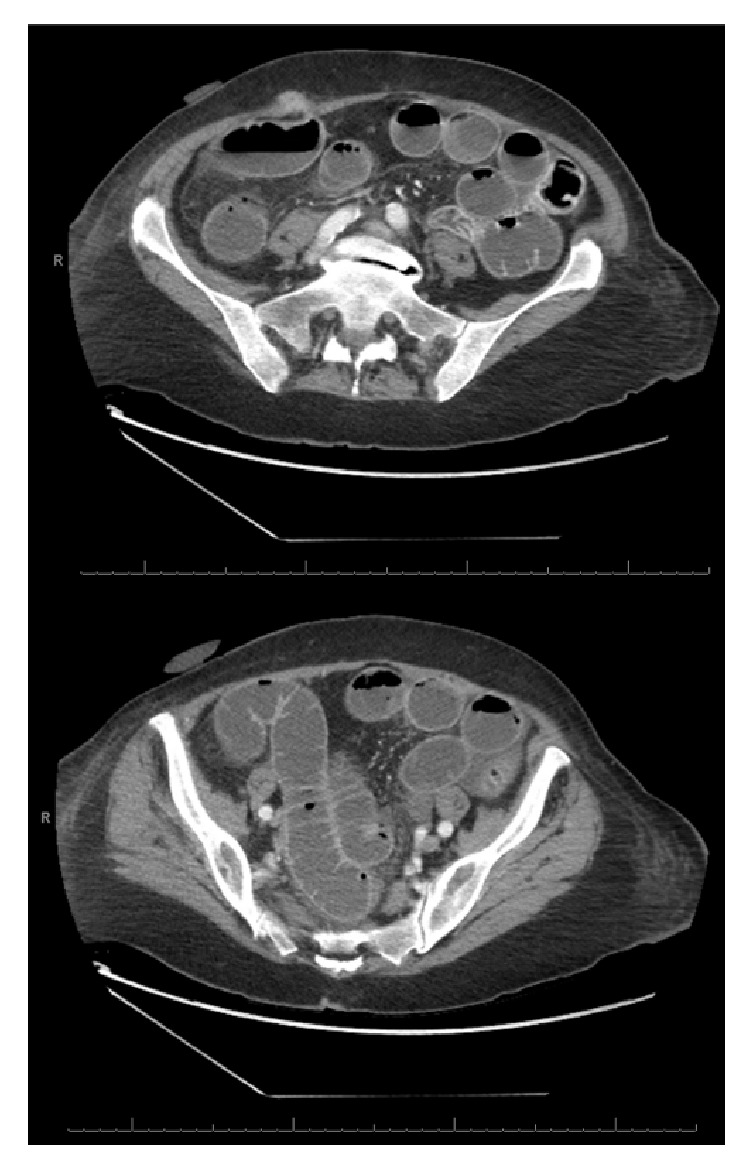
First CT scan abdomen and pelvis. Contrast-enhanced axial images showing port site hernia with diffuse small bowel dilation.

First CT scan abdomen and pelvis. Contrast-enhanced axial images showing port site hernia with diffuse small bowel dilation.