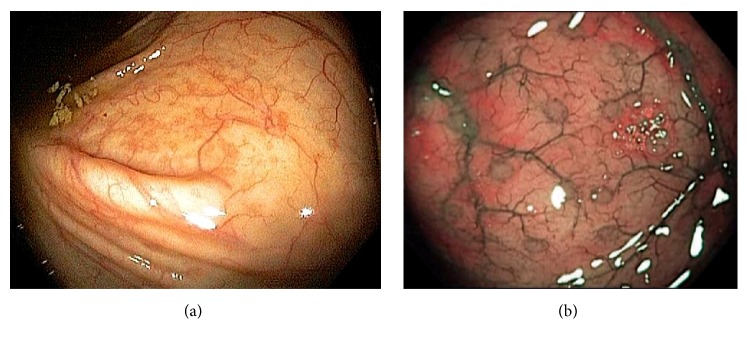
Figure 6.
Endoscopic images of the cecum. Normal vascular pattern within the ileocecum consists of branching vascular network within background of pink mucosa and intramural arteries penetrating the colonic wall (a). Gut associated lymphoid tissue is abundant in the ileocecum and they can appear as Peyer's patches, lymphoid nodules, and periappendiceal and appendiceal lymphoid follicles. These normal findings are best observed under digital chromoendoscopy (b).

