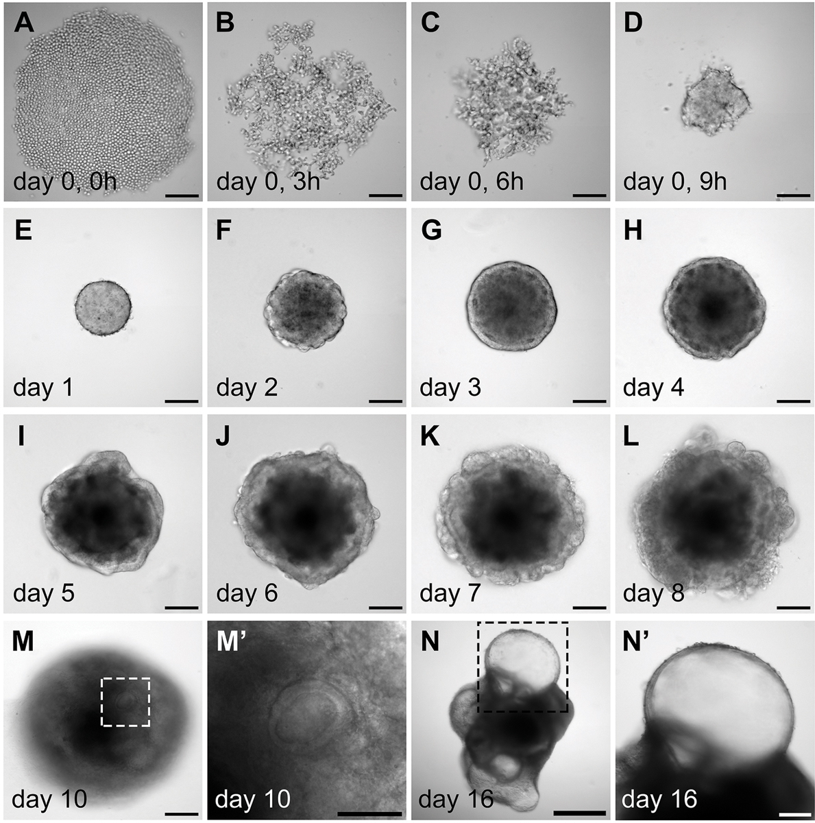
Figure 3.

Morphology of the aggregates during in vitro differentiation. (A–D) Aggregation of the mouse ES cells during day 0, which is likely mediated by cell surface adhesion proteins. (E–L) Morphological changes of the aggregates during day 1 to day 8 culture. Note that after Matrigel addition on day 1, an epithelium develops on the surface of the aggregates on day 3 (G). In a self-guided manner, some of the interior cell mass breaches out during day 6 to day 8. Concurrently, the epithelium is rearranged from the outer surface to the interior of the aggregates (J–L). (M– M’) Vesicles embedded inside the aggregates become apparent on day 10. (N–N’) A day 16 aggregate with multiple protruding vesicles. (M’) and (N’) are higher magnification images of (M) and (N), respectively. Scale bars, 100 µm (A–M, N’); 50 µm (M’); 500 µm (N).
