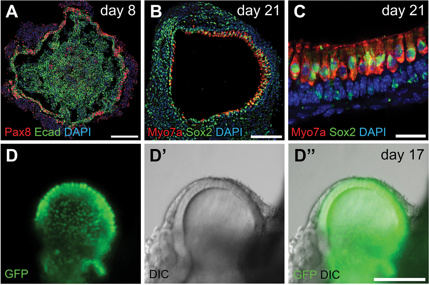
Figure 4.

Preplacodal ectoderm on a day 8 aggregate and inner ear sensory epithelium on later staged aggregates. (A) Pax8 and E-cadherin (Ecad) label the preplacodal regions on a day 8 aggregate. (B–C) Inner ear hair cells expressing Myo7a and Sox2 tightly organized at the interior surface of vesicles on day 21. (D–D”) Live Imaging of a Atoh1-nGFP aggregate. GFP with nuclear localization signal (nGFP) is expressed under an Atoh1 promoter, thus marking the inner ear hair cells. GFP signals in (D) and (D”) are overlaid from two focal planes. Scale bars, 100 µm (A–B, D–D”); 20 µm (C).
