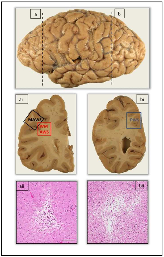
Figure 2.
Representation of the anatomic location of anterior and posterior watershed regions taken for evaluation of microscopic infarcts. The vertical dashed line (a) demarcates the level at which the anterior watershed regions, midfrontal and white matter anterior watershed regions were taken in the coronal brain slab (ai). Image aii shows the presence of a chronic microscopic infarct in the cortex of the midfrontal watershed region. The vertical dashed line (b) demarcates the level at which the posterior watershed region was taken in the coronal brain slab (bi). Image bii shows the presence of a chronic microscopic infarct in the white matter of the posterior watershed region. Abbreviations: MAWS - Midfrontal anterior watershed; WMAWS – White matter anterior watershed; PWS – Posterior watershed. Scale bar 100μm.

