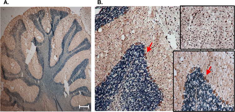
Figure 2. Localization of CCTβ in cerebellar Purkinje cells of rat brain.
Immunohistochemical analysis (magnification: X20) of 10-μm sagittal sections demonstrated a relatively high expression of CCTβ in Purkinje cells of the cerebellum, which are indicated by red arrows, with enlarged inset view (magnification: X50). Top inset (magnification: X50) indicates nuclear CCT staining throughout peripheral midbrain. Scale bar, 50 μm. Images are representative of sections analyzed from at least 3 (n = 3) separate animals.

