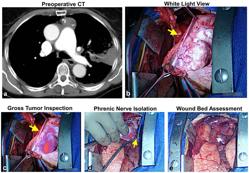
Figure 5: Fluorescent imaging provides real-time information during dissection of tumor form phrenic nerve.
In four subjects, mediastinal tumors were found in close proximity the phrenic nerve. Using fluorescent information, tumors were carefully dissected thus preserving the phrenic nerve. Data from Subject 13 is provided as a representative example. Preoperative CT (a) and intraoperative bright light views (b) of a 7.1cm thymoma. During resection, the tumor displayed high fluorescence relative to the phrenic nerve (c). Using real-time fluorescent information, the tumor was carefully dissected from the nerve (d). No residual tumor was identified after tumor resection (e).

