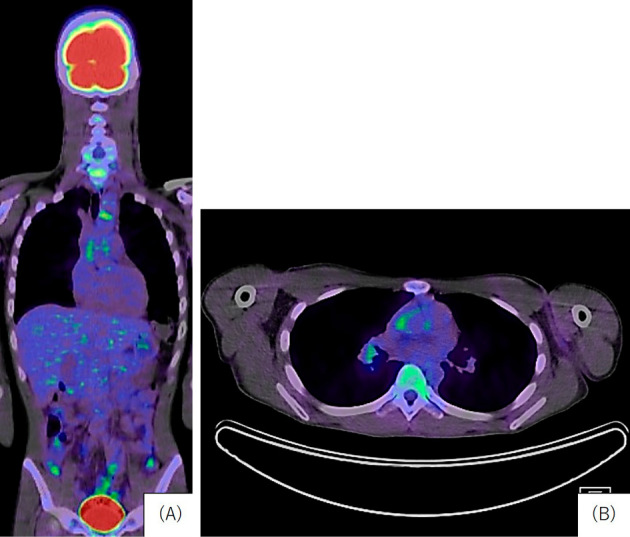
Figure 2.

>Fluoro-D-glucose positron emission tomography-computed tomography. A: A mild uptake was observed in the ascending aorta and left common carotid artery (coronal). B: A mild uptake in the right pulmonary artery and left common carotid artery (axial).
