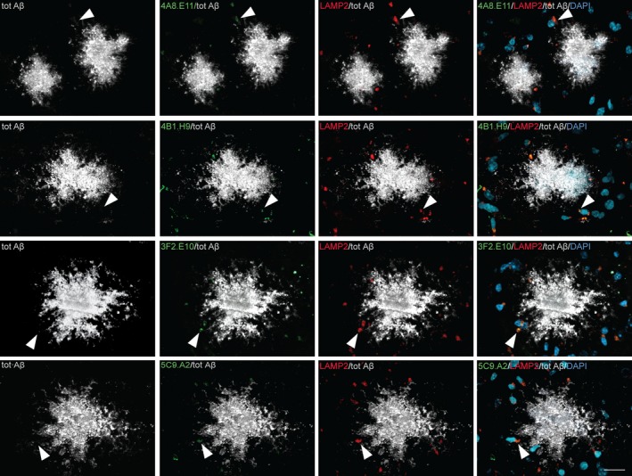
Figure 4.

mAbs reveal punctate staining around plaques but don't associate with the plaques in an AD mouse model cortex. The brain cortex obtained from APP102/TTA mice was stained to analyze the localization of our mAbs‐binding signal. Immunohistochemistry reveals that mAbs 4A8.E11, 4B1.H9, 3F2.E10, and 5C9.A2 (green) detect small, subdiffraction‐limited spot size regions surrounding the total α‐Aβ antibody‐positive plaques (grey). mAbs signal co‐localizes with the lysosome marker LAMP2 (red) localized in the vicinity of cell nuclei stained by the neuronal marker DAPI (dark cyan), but not with the plaques. Arrowheads indicate spots where co‐localization was observed; scale bar = 20 µm
