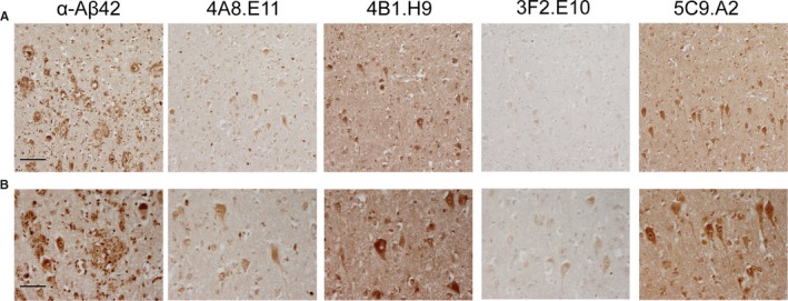
Figure 5.

Conformation‐specific mAbs stain pyramidal cells in AD human frontal cortex. The brain cortex obtained from familial AD patients was stained to analyze the localization of our mAbs‐binding signal. DAB immunohistochemistry reveals that while the α‐Aβ42 antibody stains plaques, 4A8.E11, 4B1.H9, 3F2.E10, and 5C9.A2 do not bind to plaques but specifically stain pyramidal cells. Scale bar = 500 µm in A and 250 µm in B
