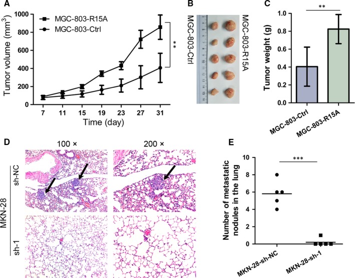
Figure 3.

RPS15A promotes GC cell growth and metastasis in vivo. A, MGC‐803‐Ctrl and MGC‐803‐RPS15A cells (2×10⁶) were injected subcutaneously into the groin of nude mice (n = 5 for either group). Tumour volumes were measured on the indicated days. Error bars represent the mean ± SD. **P < 0.01 by Student's t test. (B‐C) One month after tumour cell injection, mice were killed. The representative xenograft tumours were shown (B) and the average tumour weight was measured (C). Error bars represent the mean ± SD. **P < 0.01 by Student's t test. D, MKN‐28‐sh‐NC and MKN‐28‐sh‐1 cells (5 × 10⁶) were injected into the tail vein of nude mice (n = 5 for either group) to establish a lung metastasis model. Representative results of H&E staining of lung metastatic nodules were shown. The metastatic nodules were indicated by arrows. E, The number of lung metastatic nodules was shown. ***P < 0.001 by Student's t test
