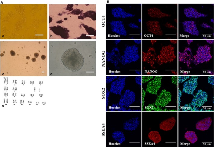
Figure 1.

Characterization of porcine‐induced pluripotent stem cells (iPSCs). A, Representative image of porcine iPSCs with well‐defined borders (a), expression of pluripotency markers alkaline phosphatase (b) and embryoid bodies (c‐d), Scale bars = 50 A). Karyotyping results of porcine iPSCs (e). Normal chromosomes (36 + XX, p26) were observed. B, The expression of pluripotent markers in porcine iPSCs as assessed by immunofluorescence analysis. Porcine iPSCs expressed pluripotent markers, including OCT4, NANOG, SOX2 and SSEA4; Scale bars = 50 µm
