Figure 6.

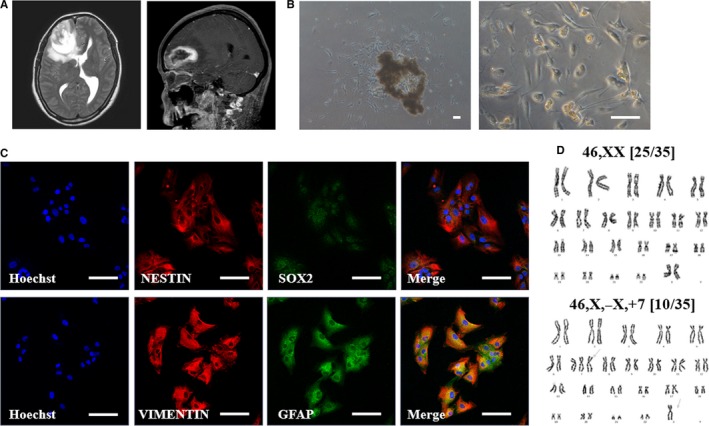
Primary culture and characterization of glioblastoma (GBM) patient‐derived cancer cell line. A, Neuroimaging of original brain tumour showed irregular shaped heterogenously enhancing intra‐axial mass in the right frontal lobe with internal haemorrhage and peritumoural oedema. B, Representative phase contrast microscopy analysis of patient‐derived primary brain tumour cells. Scale bars = 50 µm. C, Double‐immunofluorescence labelling of brain tumour cell lines. Red fluorescence labelling indicates NESTIN or VIMENTIN. Green fluorescence labelling indicates SOX2 or GFAP. Nuclei are counterstained with Hoechst (blue). Scale bars = 100 µm. D, G banded karyotype from brain cancer cell line showing chromosomal aberrations
