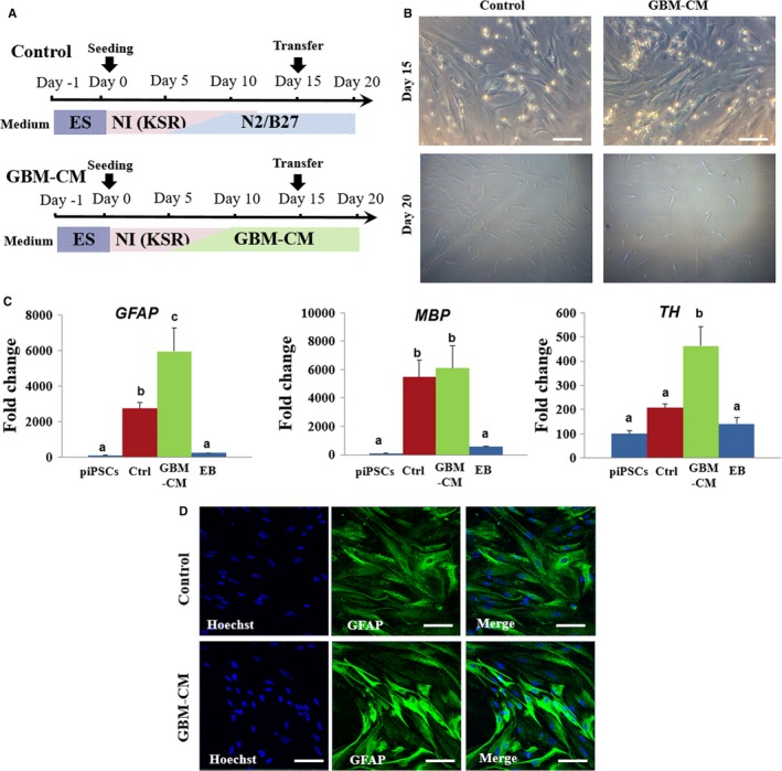
Figure 7.

Effect of GBM‐CM on development of porcine iPSC‐derived NPCs. A, Schematic representation of differentiation protocols showing treatment of GBM‐CM during neural differentiation of porcine iPSC‐derived NPCs. B, Representative image of porcine iPSC‐derived NPCs treated with or without GBM‐CM at day 20. Scale bars = 50 µm. C, Comparative real‐time PCR expression of GFAP, MBP and TH in differentiated iPSC‐derived NPCs at day 15. The expression (Mean ± SEM) was measured by comparative real‐time PCR relative to the expression in porcine iPSCs. Samples were normalized to the housekeeping gene RN18S. The experiments were performed in biological triplicates. Within the same target mRNA, values with different superscript letters (a‐c) within each column indicate significant differences between groups (P < 0.05). D, Representative images of immunofluorescence that show the increased expression of the astrocyte marker GFAP in GBM‐CM treated cells. Scale bars = 50 µm
