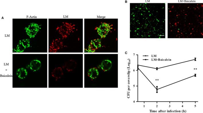
Figure 4.

Baicalein influence on intracellular bacteria at 5 h pi (A) Bacteria and F‐actin staining at 5 h after infection. B, Influence of baicalein on intracellular bacterial viability. Infection was performed as described. The lysates of infected cells at 5 h pi were stained with Syto 9 (live bacteria stain green) or propidium iodide (dead bacteria stain red); scale bar = 10 μm. C, The inhibition of baicalein on bacterial growth in RAW264.7 cells. Infected cells were lysed with 0.2% Triton‐X100 at 0.5, 2 and 5 h after infection. Intracellular bacteria were analyzed with a CFU assay. **, P < 0.01
