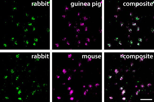
Figure 1.

Validation of VGLUT3 antibodies for immunofluorescence. Antibodies raised in rabbit, guinea pig, and mouse label the same terminal-like structures in inner lamina II in the rat spinal cord. The micrographs are single deconvolved optical sections acquired with a 63×/1.4 oil immersion objective. Scale bar, 5 µm, valid for all panels.
