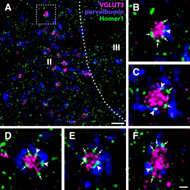
Figure 9.

Synaptic connections between VGLUT3+ C-LTMRs and parvalbumin neurons in rat dorsal horn. A, A portion of lateral Lamina IIi from a lumbar spinal cord section immunolabeled for VGLUT3, parvalbumin, and Homer1. Many VGLUT3+ terminals are apposed to parvalbumin+ processes with Homer1+ puncta. Roman numerals denote Rexed’s laminae. Dashed line indicates border between lamina II and III. Dashed frame indicates the region magnified in B. Scale bar, 5 µm. B–F, Examples at higher magnification of VGLUT3+ terminals apposed to parvalbumin processes. Arrowheads indicate Homer1+ puncta, associated with parvalbumin processes, apposed to the VGLUT3+ terminals. Arrows indicate Homer1+ puncta associated with the VGLUT3+ terminals but not with parvalbumin+ processes. Scale bar, 1 µm (B–F). All micrographs are single deconvolved optical sections obtained with a 63×/1.4 objective.
