Figure 3.
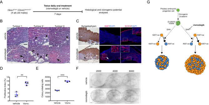
Vismodegib treatment results in higher numbers of clonogenic cells and premature formation of tumours. (A) Diagram of the experimental approach. Four-week old Hesx1 Cre/+ ;Ctnnb1 loxex3/+ male mice are dose twice daily with either vismodegib (100 mg/kg) or vehicle for 1 week, after which pituitaries are dissected and analysed both histologically and in a clonogenic assay. (B) H&E staining on FFPE histological sections of vehicle and vismodegib -treated mice showing the presence of large tumoural lesions (arrows) and cysts (arrowheads) in vismodegib-treated animals. The control pituitaries show smaller cysts (arrowheads) and no tumour lesions are detectable (n = 3 pituitaries per group). This is in agreement with our previous observations of a latency period of around 17 weeks for tumour formation (Boult et al. 2018). Scale bar: 100 μm. (C) The premature tumour lesions are Synaptophysin−ve (black arrow) by immunohistochemistry, and highly proliferative, as assessed by immnufluorescence against Ki67 (white arrow). (D) Quantitative analysis demonstrates the higher Ki67 proliferative index upon vismodegib treatment (vehicle: 3.2 ± 1.2%; vismodegib: 8.8 ± 1.0%, n = 3 pituitaries per group; P = 0.0029, Student’s t-test, vehicle = 15,213, vismodegib = 13,300 DAPI +ve nuclei counted). Scale bar: 200 μm. (E) Quantification of the clonogenic potential of pituitaries from mice treated with either vehicle or vismodegib (n = 3 pituitaries per group). Note that vismodegib treatment results in drastic increase in clonogenic potential of nearly 90% relative to the vehicle controls (vehicle: 2597 ± 98 colonies; vismodegib: 4924 ± 167 colonies, n = 3 pituitaries per group; P = 0.0003, Student’s t-test). (F) Representative examples of plates seeded with 2000, 4000 and 8000 pituitary cells from vehicle and vismodegib-treated Hesx1 Cre/+ ;Ctnnb1 loxex3/+ mice. Plates are stained with haematoxylin. (G) Schematic summary of main findings. The expression of oncogenic β-catenin in Hesx1 Cre/+ ;Ctnnb1 loxex3/+ mice results in formation of β-catenin-accumulating cell clusters, which secrete SHH and activate the pathway in surrounding tumour cells inducing a more quiescent phenotype characterised by exit of a proliferative, Ki67+ve state. vismodegib-mediated inhibition of the SHH pathway leads to more proliferative and aggressive tumours. All graph bars represent mean ± s.d.

 This work is licensed under a