
Figure 3.
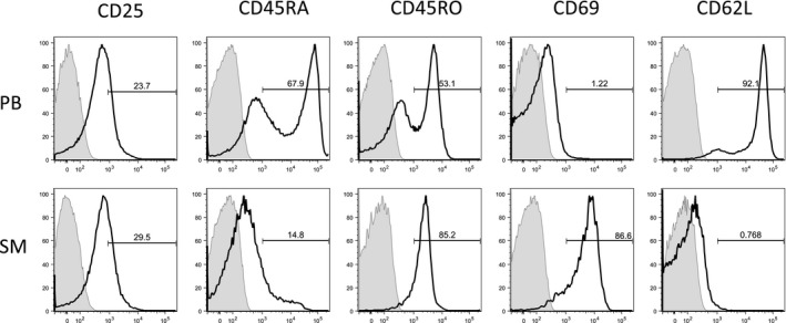
Activation status of CD4+ T cells in in peripheral blood and synovial membrane. Flow cytometry analysis of activation markers from one representative end‐stage bi‐compartmental osteoarthritis (OA) patient is shown for peripheral blood (PB) and synovial membrane (SM). Samples were stained with anti‐CD4‐fluorescein isothiocyanate (clone 2A3) to identify CD4+ cells. The cells were further stained for the following surface markers: CD25 (clone RDR5), CD45RA (clone HI100), CD45RO (clone UCHL1), CD69 (clone FN50) and CD62L (L‐selectin, clone DREG‐56). Cells stained with isotype controls are shadowed in the histograms. The bold‐type line represents the CD4+ T cells.