Figure 3.

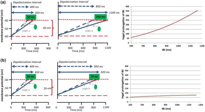
Effects of ACh release on the diastolic depolarization rate of the pacemaker cells in the SA. (a) Fixed angle scenario. The same amount of ACh release decreases the slope of diastolic depolarization by a fixed angle (α) at shorter (400 ms, left column) and longer (850 ms, middle column) diastolic depolarization intervals. This change prolongs the heart period less when the mean heart period is shorter (with faster mean diastolic depolarization of the pacemaker cells) than when mean heart period is longer (+50 ms vs. +250 ms). The graph on the right provides an illustration of this strong accumulative vagal prolongation effect across a heart period range of 600 to 1,200 ms. (b) Relative angle scenario. The same amount of ACh release decreases the slope of diastolic depolarization of the pacemaker cells by angles (α) or (β) that scale with the mean ongoing slope of diastolic depolarization. Hence, the effect on heart period is rather similar across shorter (400 ms, left column) and longer (850 ms, middle column) durations of the diastolic depolarization interval (+50 ms vs. +70 ms). The graph on the right provides an illustration of this weak vagal prolongation effect across a heart period range of 600 to 1,200 ms
Figure 3.

Effects of ACh release on the diastolic depolarization rate of the pacemaker cells in the SA. (a) Fixed angle scenario. The same amount of ACh release decreases the slope of diastolic depolarization by a fixed angle (α) at shorter (400 ms, left column) and longer (850 ms, middle column) diastolic depolarization intervals. This change prolongs the heart period less when the mean heart period is shorter (with faster mean diastolic depolarization of the pacemaker cells) than when mean heart period is longer (+50 ms vs. +250 ms). The graph on the right provides an illustration of this strong accumulative vagal prolongation effect across a heart period range of 600 to 1,200 ms. (b) Relative angle scenario. The same amount of ACh release decreases the slope of diastolic depolarization of the pacemaker cells by angles (α) or (β) that scale with the mean ongoing slope of diastolic depolarization. Hence, the effect on heart period is rather similar across shorter (400 ms, left column) and longer (850 ms, middle column) durations of the diastolic depolarization interval (+50 ms vs. +70 ms). The graph on the right provides an illustration of this weak vagal prolongation effect across a heart period range of 600 to 1,200 ms
