
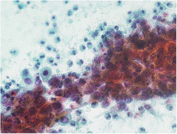
Figure 2.
Cytology of the endometrium. Atypical squamous cells revealing round to oval nuclei with coarse chromatin and prominent nucleoli. Many eosinophils are seen in the background (Pap. staining, 400×).
Official websites use .gov
A
.gov website belongs to an official
government organization in the United States.
Secure .gov websites use HTTPS
A lock (
) or https:// means you've safely
connected to the .gov website. Share sensitive
information only on official, secure websites.
Cytology of the endometrium. Atypical squamous cells revealing round to oval nuclei with coarse chromatin and prominent nucleoli. Many eosinophils are seen in the background (Pap. staining, 400×).