Figure 3.

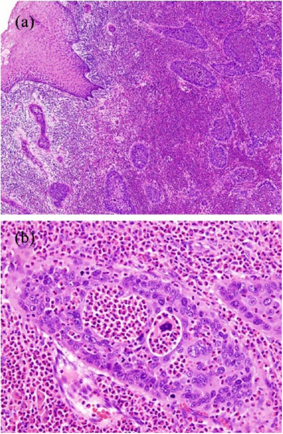
Histological findings of the uterine cervix. (a) Tumor cell nests showing stromal invasion (H&E staining, 20×).
(b) Clusters of adenocarcinomatous cells showing prominent infiltration of eosinophils (H&E staining, 200×).

Histological findings of the uterine cervix. (a) Tumor cell nests showing stromal invasion (H&E staining, 20×).
(b) Clusters of adenocarcinomatous cells showing prominent infiltration of eosinophils (H&E staining, 200×).