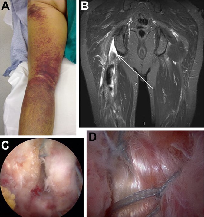
Figure 1.

(A) Clinical presentation of a proximal hamstring tear. (B) Coronal magnetic resonance imaging demonstrating a complete avulsion. (C) Endoscopic view of suture anchor placement in the anatomic footprint on the ischium with sutures passed through the tendon. (D) Final repair construct after reduction.
