Figure 1.

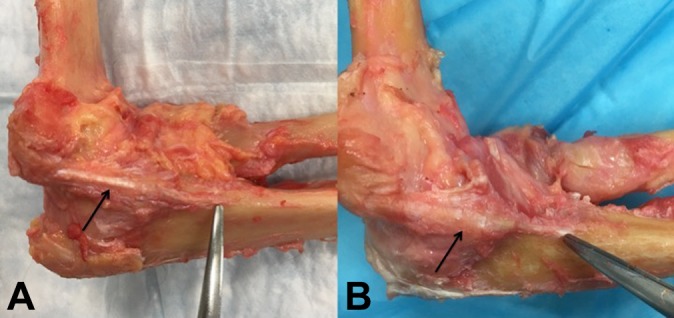
Images demonstrating the ulnar collateral ligament (UCL) in 2 of the cadaveric specimens (black arrows). Notice the distal extent of the UCL footprint on the ulna marked by the tip of the dissecting scissors in both images.

Images demonstrating the ulnar collateral ligament (UCL) in 2 of the cadaveric specimens (black arrows). Notice the distal extent of the UCL footprint on the ulna marked by the tip of the dissecting scissors in both images.