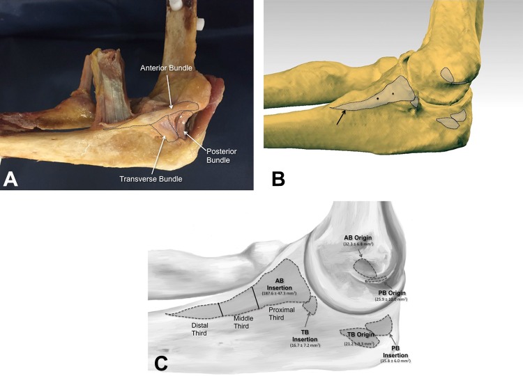
Figure 3.
(A) Image of a previously dissected cadaveric specimen with the ulnar collateral ligament (UCL) insertions of each bundle onto the ulna dotted out. (B) A computer-generated image of the origin and insertion footprints of the UCL. Notice the long insertion of the anterior bundle onto the ulna (black arrow). (C) Computer-generated image showing the insertion areas of the various bundles and how the UCL was divided into proximal, middle, and distal thirds for this study. AB, anterior bundle; PB, posterior bundle; TB, transverse bundle.

