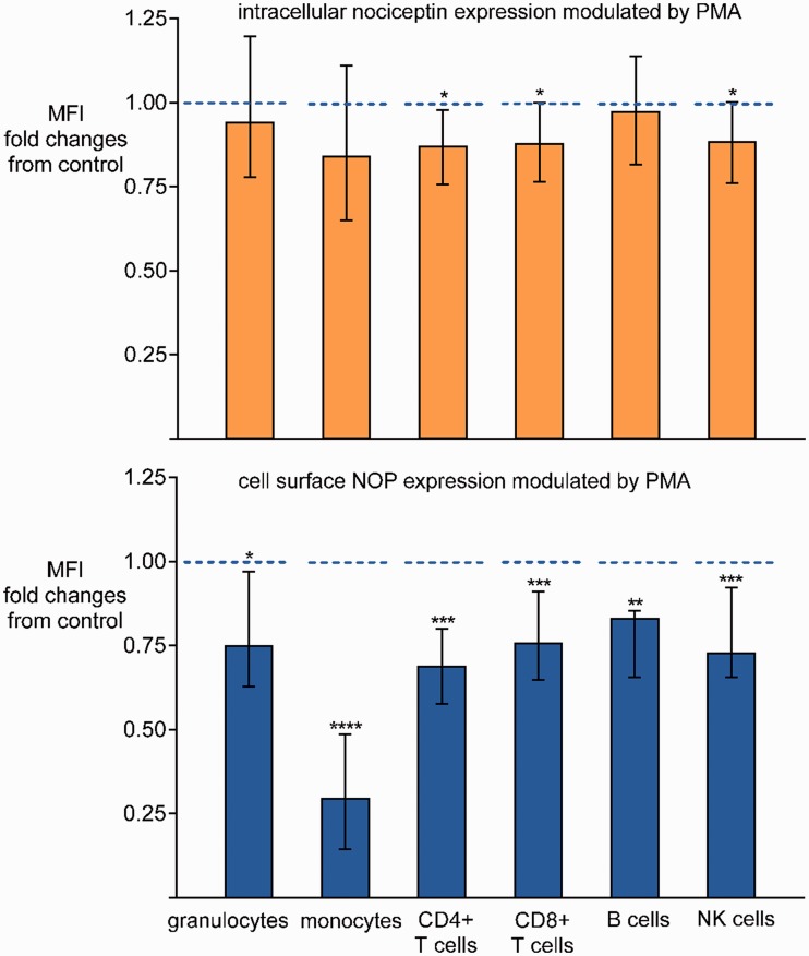
Figure 5.
Flow cytometric analysis of intracellular nociceptin and membrane NOP in blood leukocyte subsets. MFI represents the amount of intracellular nociceptin or membrane NOP in blood leukocytes. Whole blood was cultured with or without PMA 10 ng/ml for 24 h. Nociceptin and NOP protein levels in leukocyte subsets in blood samples without PMA co-incubation served as controls. Data are presented as changes in nociceptin or NOP protein levels in subsets in the PMA group relative to the respective controls . Medians and interquartile ranges, n=12 for each group. *P <0.05, **P <0.01, ***P <0.005, ****P <0.0001. PMA: phorbol-12-myristate-13-acetate; NOP: nociceptin opioid peptide receptor.

