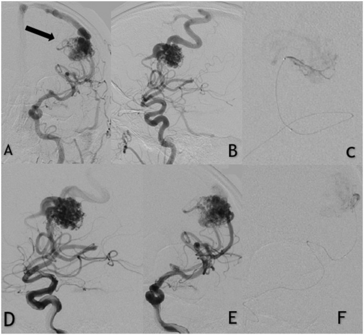
Figure 1.
Patient with unruptured left frontal arteriovenous malformation (AVM). (a and b) Anteroposterior digital subtraction angiography shows the AVM fed by the left middle cerebral artery. (d and e) Selective contrast injections in the left internal carotid artery. (c and f) Apollo microcatheter injection of the middle cerebral artery afferent demonstrating embolization of the nidus with precipitating hydrophobic injectable liquid. Note that the tip of the microcatheter is clearly visible in the cast.

