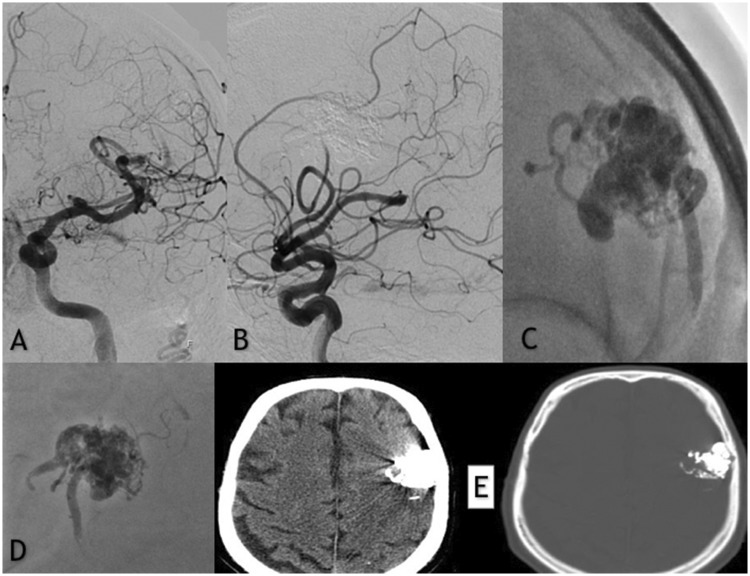
Figure 2.
(a and b) Frontal and lateral subtracted digital subtraction angiography injections showing the complete embolization of the left frontal unruptured arteriovenous malformation. (c and d) Native image demonstrating the opacities of the precipitating hydrophobic injectable liquid cast used for the embolization. (e) Axial non-contrast enhanced post-embolization computed tomography (CT) scan with minimal artifacts noted.

