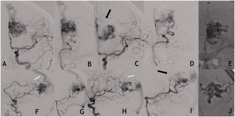
Figure 3.
(a and f) Left internal carotid and left vertebral artery (b and g) contrast injections demonstrating a left-sided unruptured occipito-parietal arteriovenous vascular malformation (AVM). Frontal (c and d) and lateral (h and i) views showing partial embolization of the AVM. Note the AVM remnant with reduced number of arterial feeders and changes in the venous drainage. (e and j) Precipitating hydrophobic injectable liquid.

