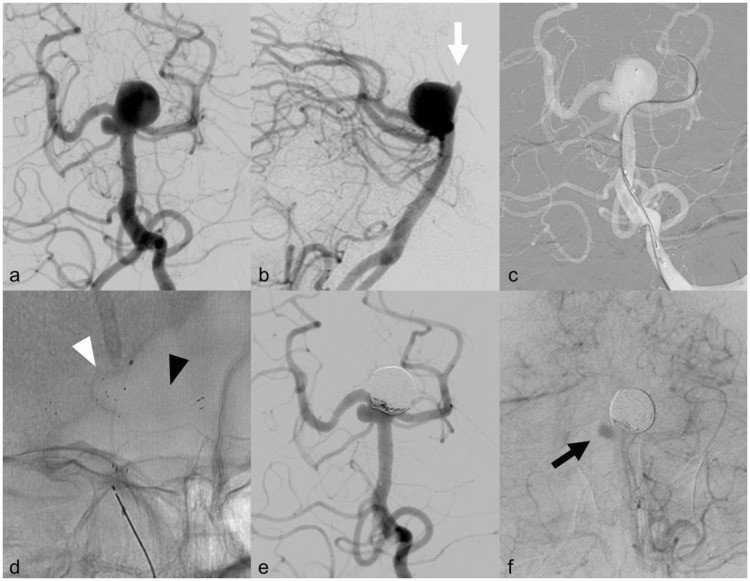
Figure 1.
Endovascular procedure. (a) Frontal view of the basilar tip aneurysm during left vertebral artery injection. Please note the small right superior cerebellar artery (SCA) aneurysm; (b) lateral view of the basilar aneurysm during left vertebral artery injection showing the antero-cranial bleb (white arrow); (c) frontal view (roadmap) during left vertebral artery injection showing the microcatheter in the left posterior cerebral artery (PCA). Please note the Barrel stent already implanted from the left P1 segment to the basilar trunk and the microcatheter inside the sac. (d) Unsubtracted frontal view of the two stents deployed (white arrowhead: Barrel vascular reconstruction device (VRD); black arrowhead: Atlas stent) and the microcatheter inside the sac; (e) frontal view of the basilar aneurysm after left vertebral artery injection at the end of the embolization showing the complete exclusion of the sac. (f) Frontal view of the right SCA aneurysm after left vertebral artery injection. Please note the contrast media stagnation inside the sac as a mild flow-diversion effect.

