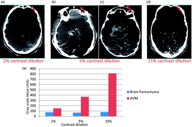
Figure 4.
Visualization and quantification of arteriovenous malformation (AVM) and brain parenchyma simultaneously using contrast-enhanced cone-beam computed tomography. The contrast medium dilution was varied from 2% to 15%. The contrast medium was injected in the vertebral artery. A slightly higher concentration of contrast medium (5%) resulted in optimal visualization of AVM and brain parenchyma because of further dilution from another vertebral artery and bony skull base.

