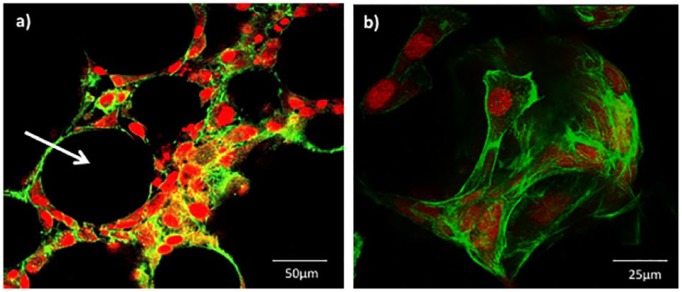
Figure 7.
Confocal laser scanning microscopy images of MG63 cells attached to Ti5 glass microspheres prepared and stained on day 13 under 150 rpm conditions. Phalloidin stain was used to identify the actin filaments of the cytoskeleton (green) while the propidium iodide (PI) stain was used for the nuclear staining (red). Image (a) provides a sliced cross-section through a cluster of microspheres cultured with MG63 cells, while image (b) illustrates a 3D rendered image of an individual microsphere. Scale bar is (a) 50 µm and (b) 25 µm, respectively.

