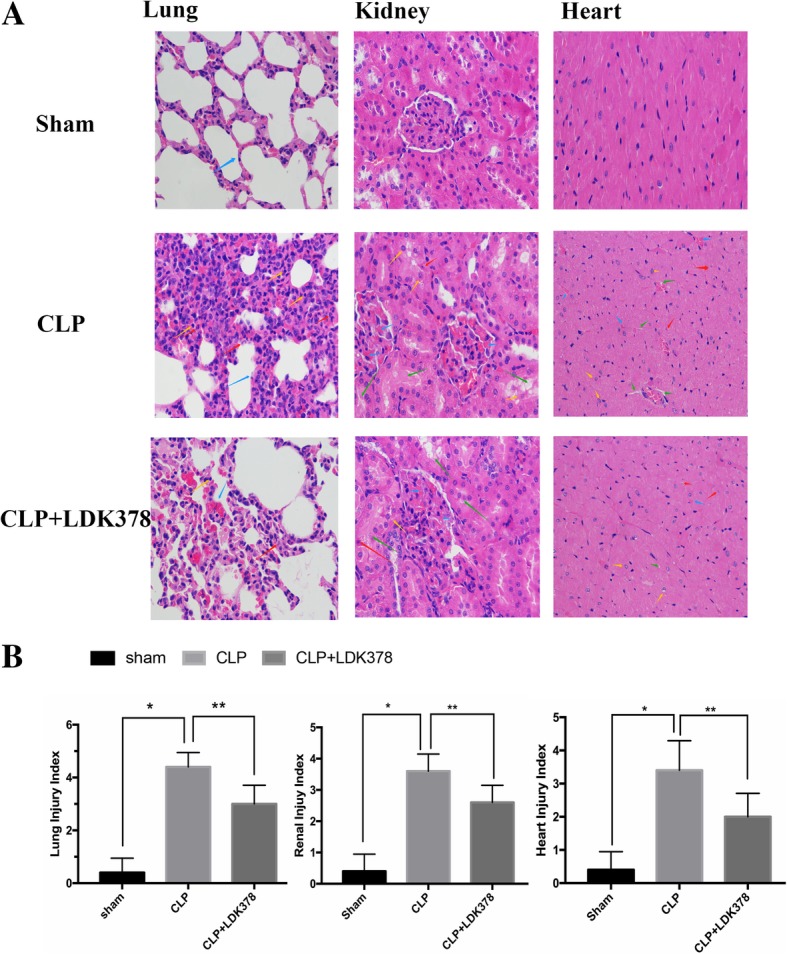
Fig. 3.

Morphological changes in major organ tissue pathology (400X). (a) Representative hematoxylin and eosin staining results for lung and kidney sections in rats. (b) Semi-quantitative analysis of H&E staining. *p < 0.05 vs sham group; #p < 0.05 vs CLP group. Lung: Inflammatory cells were diffusely infiltrated, and the alveolar wall was significantly widened in CLP group; a smaller number of inflammatory cells infiltrated in the LDK378 treatment group, and the alveolar wall was slightly widened, individual inflammatory cells infiltrated in Sham group, and no obvious broadening of the alveolar wall was observed (red arrow: neutrophils; yellow arrow: lymphocytes; blue arrow: alveolar wall). Kidney: Hyaline degeneration of renal tubular epithelial cell, necrosis, shedding and hemorrhage in CLP group; Lesions were also seen in the LDK378 treatment group, but range was smaller than CLP group. (yellow arrow: hyaline degeneration: red arrow, necrosis; green arrow: shedding; blue arrow: hemorrhage). Heart: Increased cell gap, necrosis, hemorrhage was seen in CLP group; the number of necrosis and hemorrhage were smaller in LDK378 treatment group (green arrow: cell gap increased; yellow arrow: Cell degeneration; red arrow: necrosis; blue arrow: hemorrhage)
