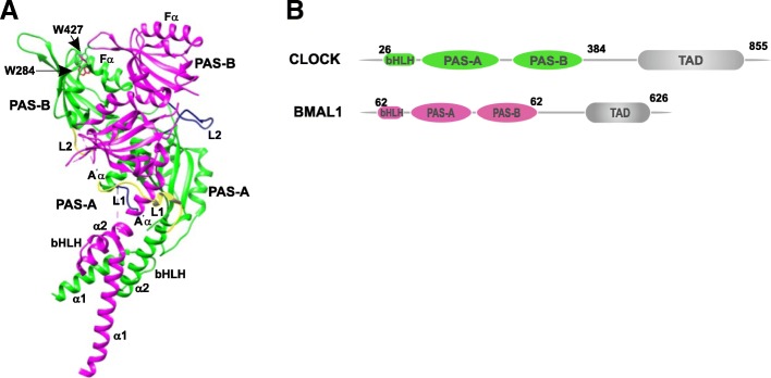
Fig. 10.
Crystal structure of the mouse CLOCK–BMAL1 complex (PDB 4F3L). a The ribbon diagram of the complex shows the CLOCK subunit in green and BMAL1 in pink. Yellow and blue highlight the respective linker regions between the domains. b Domain architecture of CLOCK and BMAL1 depicting the basic helix-loop-helix domain and the two PAS domains

