
Fig. 6.
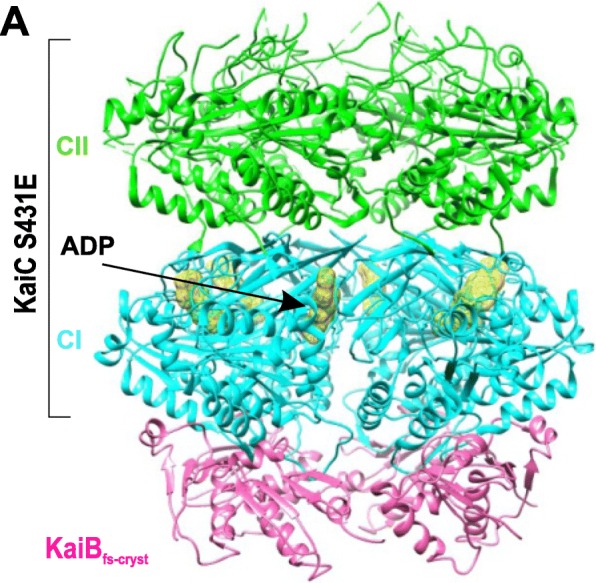
Kai clock protein complex assembly. a A 3.87-Å structure of KaiBfs-cryst*and KaiC S431E complex hexamer (PDB 5JWQ) with KaiBfs-cryst* in hot pink, the KaiC CI domain ring in cyan, CII in green, and ADP densities in yellow
Official websites use .gov
A
.gov website belongs to an official
government organization in the United States.
Secure .gov websites use HTTPS
A lock (
) or https:// means you've safely
connected to the .gov website. Share sensitive
information only on official, secure websites.
Kai clock protein complex assembly. a A 3.87-Å structure of KaiBfs-cryst*and KaiC S431E complex hexamer (PDB 5JWQ) with KaiBfs-cryst* in hot pink, the KaiC CI domain ring in cyan, CII in green, and ADP densities in yellow