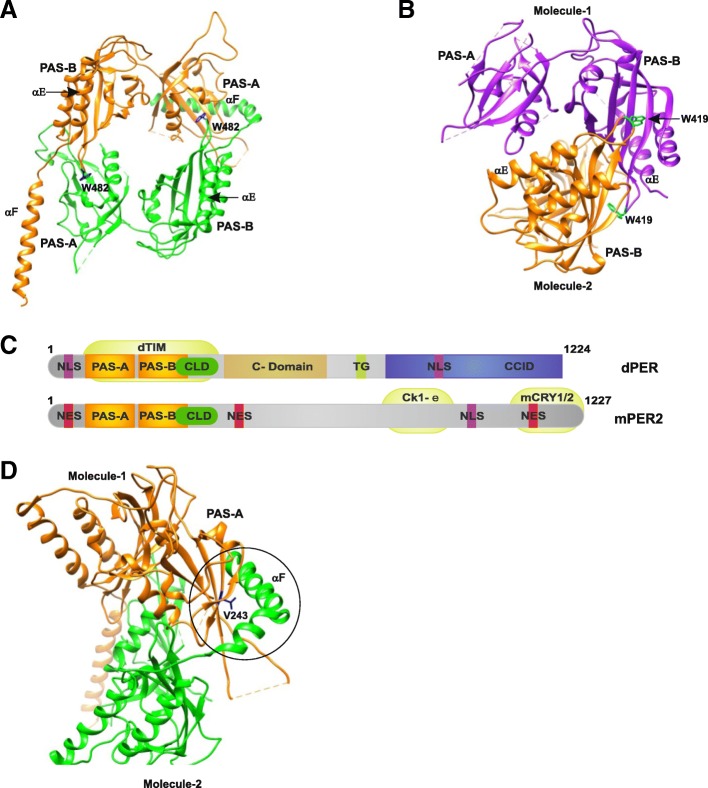
Fig. 8.
Crystal structures of the period proteins. a dPER (PDB 1WA9) and b mPER2 (PDB 3GDI) dimers in cartoon representation. The conserved Trp482 (dPER, dark blue) and Trp419 (mPER2, cyan) residues are shown in stick representation. c The domain architecture of dPER and mPER2 proteins. The two PAS domains (PAS-A and PAS-B), the cytoplasmic localization domain (CLD, green), the conserved C-domain (light brown), nuclear localization signals (NLS, purple), NES (red), the threonine-glycine (TG) repeat region, and the dCLK:CYC inhibition domain (CCID, blue) of dPER and/or mPER2 are shown. CKIe, mCRY1/2, and dTIM are shown at their binding sites. d dPER structure representing the PAS-A–αF interaction (encircled region) interface and depicting the location of V243 (blue)

