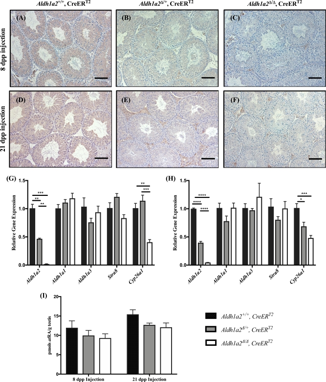
Figure 2.
Elimination of Aldh1a2 using the inducible CreERT2. Analysis of Aldh1a2+/+, CreERT2; Aldh1a2▵/+, CreERT2; and Aldh1a2▵/▵, CreERT2 animals injected with tamoxifen. Representative immunohistochemistry from animals injected at 8 (A, B, and C; 68 dpp at euthanasia) and 21 dpp (D, E and F; 81 dpp at euthanasia). Cross-sections are stained for ALDH1A2 protein and immunopositive cells are indicated by brown precipitate. qRT-PCR analysis was performed to determine the relative expression of Aldh1a2, Aldh1a1, Aldh1a3, Stra8, and Cyp26a1 in animals injected with tamoxifen at 8 (G) and 21 dpp (H). (I) Graphical representation of atRA measurements. Scale bars = 100 μm. n = 3–7. *P < 0.05, **P < 0.01, ***P < 0.001, and ****P < 0.0001.

