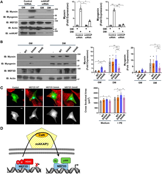
Figure 8.
Regulation of myocyte phenotype by MEF2D Ser-444 phosphorylation. A, C2C12 cells transfected with mAKAP or control siRNA were cultured for 24 h in GM and DM before analysis by Western blotting. p (two-way ANOVA for both factors and interaction) < 0.03 for both genes. B, same as in A except with cells expressing GFP- and FLAG-tagged MEF2D WT and mutant proteins. p (two-way ANOVA for MEF2D mutants and interaction) < 0.01 for myosin; p (two-way ANOVA for MEF2D mutants) = 0.02 for myogenin. C, neonatal rat ventricular myocytes were transfected with plasmids expressing FLAG-tagged MEF2D mutants and marker GFP and stained with α-actinin (red) and FLAG (blue) antibodies. The GFP channel is shown also in grayscale below. Bar, 10 μm. Myocyte cross-section areas measured using GFP images are shown. n = 3 independent experiments for A–C. p (two-way ANOVA for both factors) < 0.05. D, model for regulation of MEF2D chromatin complexes by perinuclear mAKAPβ signalosomes. We propose that MEF2D dynamically associates with chromatin, such that transient association with mAKAPβ facilitates its regulation by CaN. HDAC nuclear export is also promoted by mAKAPβ-dependent signaling (12).

