
Figure 4.
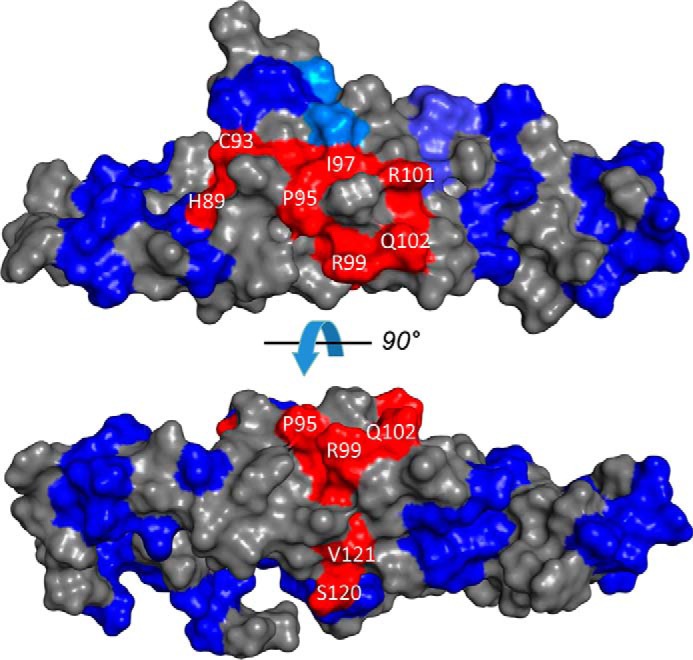
The RetGC interface localizes to the central part of the RD3 surface with a similar view as in Fig. 3C rotated 90° counterclockwise. Fragments of RD3 primary structure implicated in the inhibitory binding of RD3 to RetGC1 (red) and those that sustain mutagenesis without loss of the inhibitory binding to the cyclase (blue) (14) are superimposed on the RD3 NMR model.