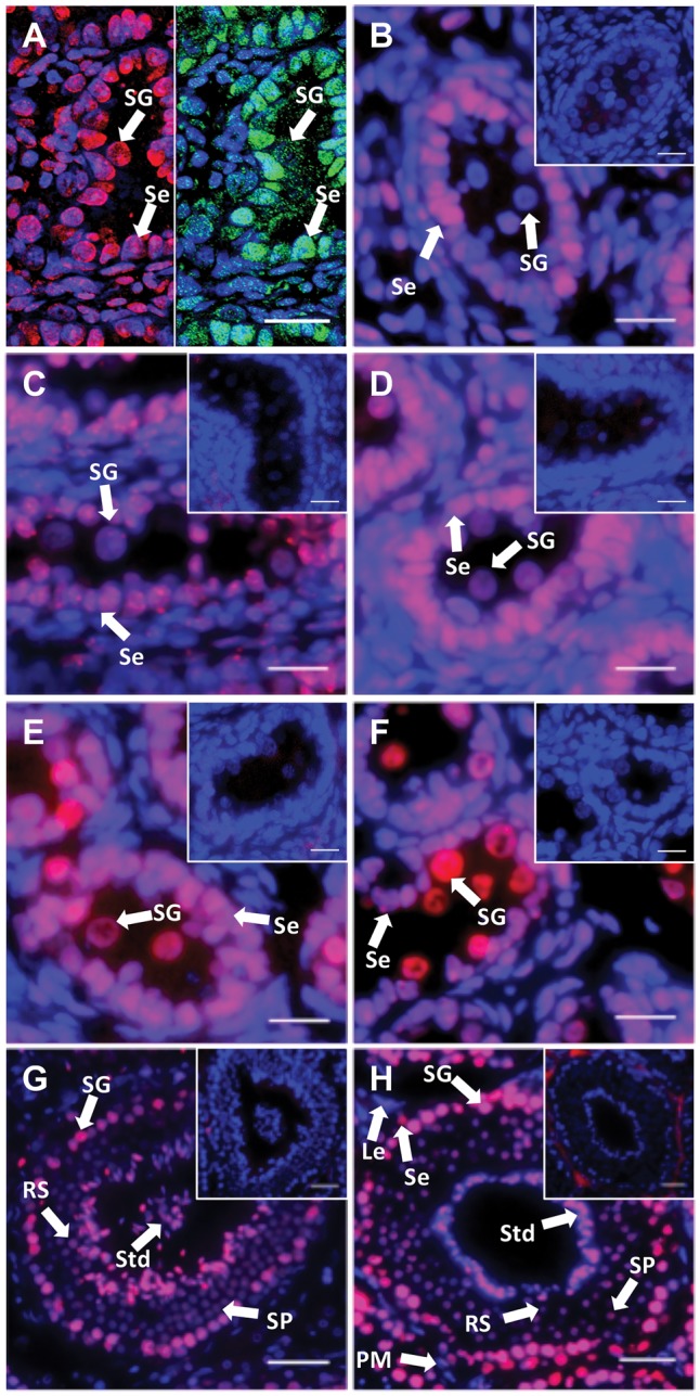
Figure 1.

5 mC localization during spermatogenesis. DNA methylation was investigated by 5 mC immunohistochemical staining. DAPI and 5 mC stain nuclei blue and red, respectively (grey and white in black and white (BW) respectively). (A) Day 10 pp. Left-hand side: 5 mC staining (red); right-hand side SOX9 staining (green). (B) Day 25 pp. (C) Day 50 pp. (D) Day 70 pp. (E) Day 100 pp. (F) Day 200 pp. (G) and (H) Adult. All insets are negative controls. (A) Scale bar 8 μm. (B–F) Scale bars 20 μm. (G and H) Scale bars 50 μm. Abbreviations: Le, Leydig cells; PM, peritubular myoid cells; RS, Round spermatids; Se, Sertoli cells; SG, spermatogonia; SP, spermatocyte; Std, spermatid.
