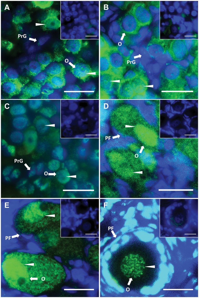
Figure 7.
DNMT3L localization during oogenesis. Immunofluorescent localization of tammar DNMT3L protein in developing and adult ovaries. DAPI stains nuclei blue (grey in BW) and DNMT3L is stained green (white in BW). Arrow heads represent nuclear localization. (A) Day 25 pp. (B) Day 50 pp. (C) Day 70 pp. (D) Day 110 pp. (E) Day 200 pp. (F) Adult. All insets are negative controls. Scale bars 20 μm. Abbreviations: O, oogonia; PrG, pre-granulosa cells; PF, primary follicles.

