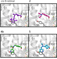
Figure 3.

In silico modeling. Predicted binding modes of ALDH1A1 substrates from in silico docking studies. Predicted binding modes in ALDH1A1 of 9‐cis‐retinal (purple), 2 (magenta), 4 b (green) and 5 (cyan), shown in ball and stick representation. Catalytic residue C302, and Y296 which was identified in π‐stacking interactions with the substrates are shown as dark grey sticks. Ribbons are shown in grey and faded for clarity, the cofactor NAD(H) is shown in pastel orange, green, red and blue ball and stick representation.
