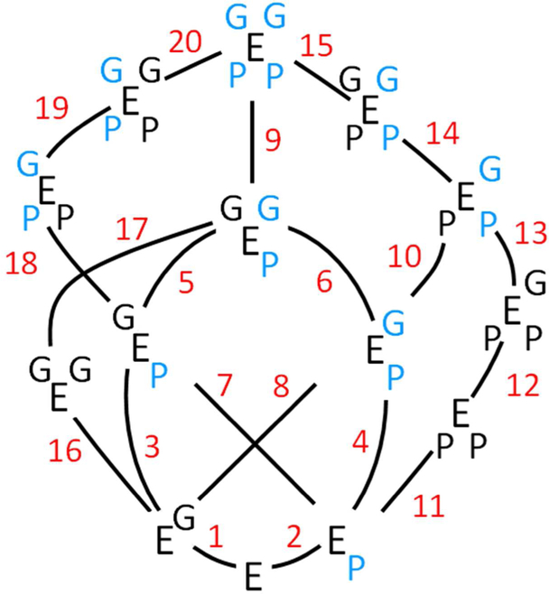
Figure 7. The Complete Interaction Scheme.
The fourteen enzyme forms involved in the bindii and interactions of EGCG (G) and PAP (P) are shown. The enzyme symbol, E, is “sided” - the two right- and left-hand corners represent binding sites on separate subunits of the SULT1A1 dimer. Upper and lower corners represent the G- and P-binding sites, respectively. The 20 binding steps that interconvert the species are numbered, and the corresponding equilibrium constants can be found in Table S1. A blue letter indicates that that ligand is bound to a subunil whose cap is closed.

